Welcome to your Digital Campus Hematology

Learn hematology where it matters: at the specimen

Learn hematology where it matters: at the specimen
Recognizing a cell is one thing. Understanding what it means in a blood or bone marrow specimen takes practice. The Digital Campus Hematology brings together a collection unmatched in its scope—more than 700 interactive, expert-annotated specimens—and over 32 hours of teaching by leading physicians. Learn from their experience, then examine the findings for yourself.
Video courses and interactive specimens included in one annual membership

Learn from experts. Then take a closer look.

The campus combines on-demand teaching with independent practice. Watch a course to understand a method or finding, then explore digital specimens at your own pace. The courses and collection support each other, whether you are learning the foundations or revisiting a difficult diagnosis.

Learn from recorded courses

Learn from senior physicians and specialists in hematology, pathology, and advanced diagnostics. Their on-demand courses take you from the foundations of microscopy to complex findings and diagnostic methods. Pause, revisit, and learn at your own pace.

Examine the whole specimen

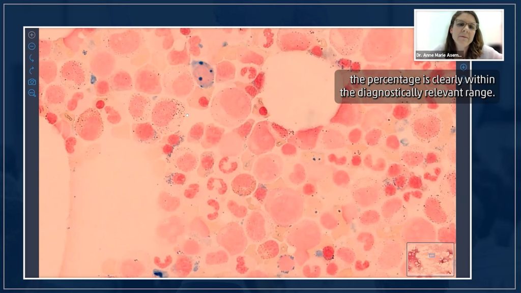
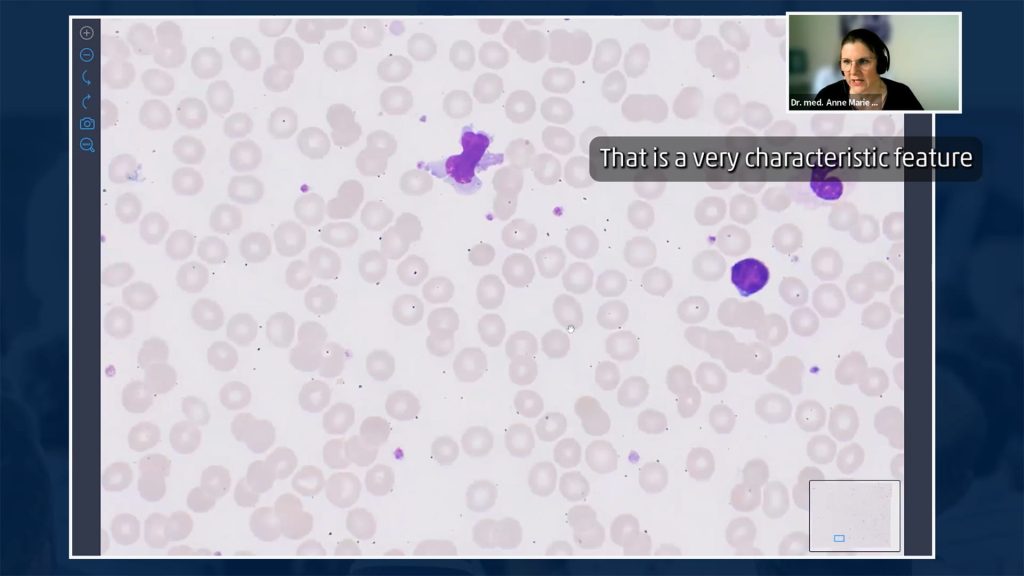
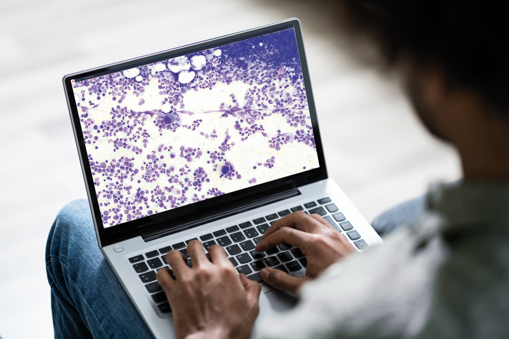
Navigate digitized peripheral blood and bone marrow smears, CSF, and other samples. Zoom in and out, measure structures, rotate the image, or use a grid. Expert annotations point to relevant findings without limiting where you can look.

Test your understanding

Use clinical cases, focused learning modules, and quizzes to practice identifying findings and checking what you have learned.

More than 700 specimens to explore beyond the textbook view

The collection covers normal morphology, common diseases, and rare findings in benign and malignant hematology. You can follow an annotation to a particular cell or structure, examine the surrounding area, and decide what you see before reading the expert’s explanation.

Anne Marie Asemissen, M.D., built this collection and annotated every case on the campus. Its breadth gives learners access to normal morphology, common diseases, and rare findings that few people could encounter during training in a single laboratory. Her annotations guide your eye, while the interactive viewer leaves the whole specimen open for your own exploration.

From recognizing a finding to putting it in context

Develop a structured approach to peripheral blood and bone marrow morphology. Recognize characteristic cytological findings, encounter rare cases, and learn where cytomorphology provides answers—and where further investigation is needed.

The courses also explain how methods such as flow cytometry, molecular genetics, and cytogenetics help clarify morphological findings.

Wherever you are in your learning journey

Whether you are taking your first steps in hematological microscopy or refining skills you use every day, the campus gives you room to learn and practice. It is set up for hematologists, physicians in training, laboratory professionals, and students interested in morphology.

For hospitals and laboratories, the campus provides specialist teaching and extensive specimen collection for staff year-round. Teams can learn and revisit cases on site, reducing the need to travel for every training opportunity.

Choose the membership that fits how you learn or teach:

Expertise you can learn from.

Dr. Anne Marie Asemissen is the driving force behind the campus’s digital specimen collection. She built and annotated the cases that make independent exploration possible.

The recorded video courses feature physicians with expertise in hematology, pathology, and specialized diagnostics. Their teaching helps you connect morphological observations with the wider diagnostic picture.

Make the next finding one you recognize

Explore the courses and specimens whenever you have time to learn. Return to a slide, compare details, and build an understanding that extends beyond a single image.

Your membership gives you access to the video courses and interactive specimens for one year.